Introduction
The human mouth is an active, complex environment where changes can happen gradually over time. From the tongue and gums to the inner cheeks, roof of the mouth, and underlying jaw structures, the soft tissues of the oral cavity are exposed daily to thermal, mechanical, and chemical stimuli. While many minor mouth sores, bumps, or discolored patches clear up on their own, persistent tissue changes require professional evaluation.Regular dental visits provide an ongoing opportunity for healthcare professionals to observe oral health and perform clinical examinations. Clinical evaluations aim to identify suspicious tissue abnormalities long before they cause significant discomfort or become noticeable to the patient. However, finding a white patch, red spot, or small ulcer during a checkup does not automatically mean a person has oral cancer; the vast majority of oral lesions turn out to be benign conditions, such as friction injuries, aphthous ulcers, or localized infections.Resources like BESTDENTALHOSPITALS help patients research dental facilities, understand standard oral health categories, and formulate meaningful questions before booking a clinical examination.
What Is Oral Cancer Screening?
An oral cancer screening is a non-invasive visual and tactile examination of the oral cavity, head, and neck performed by a dentist or healthcare professional. Its primary goal is to look for abnormal tissues, persistent lesions, or structural changes that require monitoring, treatment, or further diagnostic testing.
┌─────────────────────────────────────────────────────────────────┐
│                  ANATOMICAL ZONES EXAMINED                      │
├─────────────────────────────────────────────────────────────────┤
│ • LIPS & VERMILION BORDER  (Outer surfaces and inner lining)    │
│ • BUCCOSMUCOSA             (Inner linings of the cheeks)        │
│ • HARD & SOFT PALATE       (Roof of the mouth)                  │
│ • TONGUE                   (Dorsal, lateral borders, and base)  │
│ • FLOOR OF THE MOUTH       (Area beneath the tongue)            │
│ • GINGIVA                  (Gums and alveolar tissue)           │
│ • EXTRAORAL HEAD & NECK    (Jawline, neck muscles, lymph nodes) │
└─────────────────────────────────────────────────────────────────┘
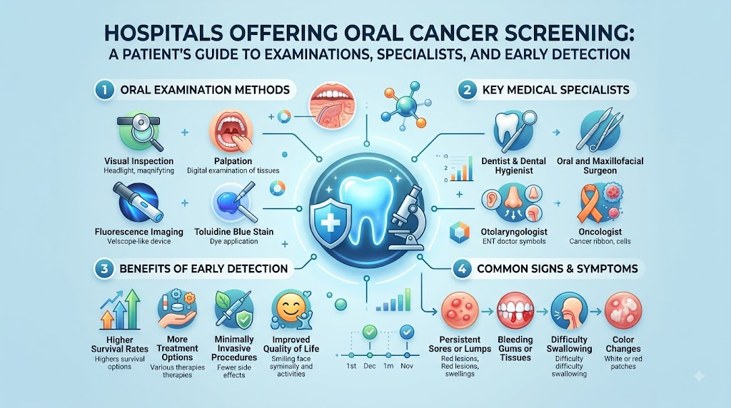
Key Components of an Oral Examination
- Patient History Review: Collecting updated details regarding medical conditions, past dental procedures, alcohol and tobacco exposure, and any newly noticed oral symptoms.
- Visual Inspection: Examining all visible oral mucosa under bright, clear light to identify changes in color, texture, surface integrity, or symmetry.
- Physical Palpation: Gently feeling the soft tissues of the mouth, floor of the jaw, and neck lymph nodes to check for subtle firm areas, swellings, or deep tissue lumps.
Important Distinction: Screening vs. Diagnosis
A clinical oral examination does not confirm or diagnose cancer by itself. Instead, screening serves as a preliminary evaluation to identify suspicious abnormalities that warrant further follow-up, monitoring, specialist evaluation, or diagnostic biopsy.
Why Is Oral Cancer Screening Important?
Oral mucosal changes often begin silently. In their initial stages, early lesions or potentially malignant disorders may cause little to no discomfort, making them easy to overlook during daily activities like brushing or eating.
                  ┌──────────────────────────────────────────┐
                  │   Core Objectives of Clinical Evaluation │
                  └─────────────────────┬────────────────────┘
                                        │
      ┌──────────────────┬──────────────┴───────────────┬──────────────────┐
      ▼                  ▼                              ▼                  ▼
┌───────────┐    ┌──────────────┐              ┌───────────────┐    ┌──────────────┐
│ Early     │    │ Timely       │              │ Symptom       │    │ High-Risk    │
│ Detection │    │ Referral     │              │ Monitoring    │    │ Focus        │
└───────────┘    └──────────────┘              └───────────────┘    └──────────────┘
- Spotting Unnoticed Changes: Regular clinical checks help catch subtle tissue modifications that patients might not see or feel.
- Promoting Timely Care: Identifying persistent lesions early allows clinicians to schedule follow-up appointments or arrange specialist referrals before a condition progresses.
- Tracking Tissue Behavior: Routine evaluations allow dentists to monitor long-standing, non-cancerous conditions (such as lichen planus or traumatic ulcers) to ensure they heal as expected.
- Focused Attention for At-Risk Patients: Individuals with higher risk profiles—such as long-term tobacco or heavy alcohol users—benefit from routine professional checks.
Note on Prevention: A clinical examination does not prevent cancer from developing. However, regular evaluations help ensure that suspicious mucosal changes receive prompt professional attention rather than remaining unnoticed.
What Happens During an Oral Cancer Examination?
An oral cancer examination is typically brief, painless, and integrated into a routine dental checkup. Understanding each phase can help patients feel comfortable and prepared.
┌────────────────────────────────────────────────────────────────────────┐
│                   Clinical Oral Examination Steps                      │
├────────────────────────────────────────────────────────────────────────┤
│ Step 1: Medical, Social, and Dental History Review                     │
│ Step 2: Extraoral Examination (Face, Jaw, Lips, and Neck Lymph Nodes)  │
│ Step 3: Intraoral Visual Inspection (Cheeks, Gums, Tongue, & Palate)   │
│ Step 4: Intraoral Palpation (Bimanual feeling of soft tissues)         │
│ Step 5: Assessment & Recommendation (Monitoring, Referral, or Biopsy)  │
└────────────────────────────────────────────────────────────────────────┘
Step 1: Medical and Dental History
The clinician reviews your general health, past medical treatments, family history, and personal risk factors. They will ask about:
- Tobacco use (cigarettes, cigars, pipes, or smokeless tobacco).
- Alcohol consumption habits.
- Betel quid or paan usage.
- Any persistent mouth sores, throat tightness, or changes in voice.
Step 2: Extraoral Examination
The examiner inspects the facial structures, lips, symmetry of the jaw, and neck area. They gently palpate (feel) the lymph nodes along the neck, under the chin, and around the jawline to check for swelling, tenderness, or fixed lumps.
Step 3: Intraoral Visual Inspection
Using a dental mirror and focused light, the clinician examines all internal surfaces of the mouth:
- Cheeks and Lips: Checking the inner lining for color changes or sores.
- Gums (Gingiva): Inspecting tissue around the teeth.
- Tongue: Examining the top surface, pulling the tongue gently to the side to check its lateral borders, and viewing the underside.
- Floor of Mouth & Palate: Inspecting the area beneath the tongue and the roof of the mouth.
Step 4: Intraoral Palpation
Using gloved fingers, the clinician gently feels the tongue, floor of the mouth, and cheeks to check for underlying firmness, thickened bands, or concealed masses.
Step 5: Next Steps & Recommendation
If all tissues look healthy, no further action is required until your next routine visit. If a non-healing or suspicious area is found, the clinician will document the finding, recommend follow-up monitoring, order further evaluation, or refer you to a specialist.
Common Warning Signs of Oral Health Changes
While many oral conditions cause temporary symptoms, persistent abnormalities should be evaluated by a dental professional.
[ Persistent Mouth Sore ] ───> Sore or ulcer that does not heal within 2 weeks
[ Discolored Patches ] ──────> Distinct white (Leukoplakia) or red (Erythroplakia) areas
[ Unexplained Lumps ] ───────> Thickened tissue, lump, or hard mass in mouth or neck
[ Sensory/Motor Changes ] ──> Numbness, persistent pain, or difficulty swallowing/chewing
Symptoms to Monitor:
- A sore or ulcer on the lip, gums, or inside the mouth that does not heal within 14 days.
- A persistent white patch (Leukoplakia) or red patch (Erythroplakia) on the oral lining.
- A unexplained lump, swelling, or thickened area on the tongue, gums, or cheek.
- Persistent pain or tenderness in the mouth, tongue, or ear.
- Difficulty or discomfort when chewing, swallowing, speaking, or moving the jaw or tongue.
- Unexplained numbness or loss of feeling in the oral tissues or lips.
- Persistent hoarseness or the feeling of something caught in the throat.
- Dentures that suddenly fit poorly or feel uncomfortable due to tissue changes.
- An unexplained, painless lump or swollen lymph node in the neck.
Clinical Guidance: According to the National Institute of Dental and Craniofacial Research (NIDCR), any sore, patch, swelling, or swallowing difficulty that persists for more than two weeks should be evaluated by a dentist or medical doctor. Most turn out to be benign, but prompt evaluation ensures accurate care.
Understanding White and Red Oral Patches
During an intraoral visual inspection, discolored patches are among the most common findings evaluated by clinicians.
┌─────────────────────────────────────────────────────────────────────────┐
│                    Leukoplakia vs. Erythroplakia                        │
├───────────────────────────────────┬─────────────────────────────────────┤
│ Feature                           │ Details                             │
├───────────────────────────────────┼─────────────────────────────────────┤
│ Leukoplakia (White Patch)         │ • Adherent white patch that cannot  │
│                                   │   be wiped off with gauze           │
│                                   │ • Often caused by mechanical        │
│                                   │   friction, tobacco, or irritation  │
│                                   │ • Requires monitoring or biopsy     │
├───────────────────────────────────┼─────────────────────────────────────┤
│ Erythroplakia (Red Patch)         │ • Smooth or velvety red area on     │
│                                   │   the oral mucosa                   │
│                                   │ • Less common than white patches    │
│                                   │   but carries a higher clinical     │
│                                   │   concern                           │
│                                   │ • Warrants prompt specialist review │
└───────────────────────────────────┴─────────────────────────────────────┘
- Leukoplakia: Refers to a predominant white patch of the oral mucosa that cannot be wiped away and cannot be clinically diagnosed as any other specific disease. While many cases are hyperkeratotic responses to local irritation, persistent white patches require clinical assessment and potential biopsy.
- Erythroplakia: Refers to a bright red, velvety patch on the oral tissues that cannot be attributed to trauma or inflammation alone. Clinical guidelines emphasize that persistent red patches carry a high association with tissue dysplasia and require prompt evaluation.
Patients should never try to scrape, cut, or self-treat discolored patches at home.
Major Oral Cancer Risk Factors
While oral cancer can occasionally develop in individuals without clear risk factors, certain environmental, lifestyle, and biological factors significantly increase clinical risk.
                  ┌──────────────────────────────────────────┐
                  │    Primary Oral Cancer Risk Factors      │
                  └─────────────────────┬────────────────────┘
                                        │
      ┌──────────────────┬──────────────┴───────────────┬──────────────────┐
      ▼                  ▼                              ▼                  ▼
┌───────────┐    ┌──────────────┐              ┌───────────────┐    ┌──────────────┐
│ Tobacco   │    │ Heavy Alcohol│              │ Betel Quid /  │    │ High-Risk    │
│ Products  │    │ Consumption  │              │ Paan Chewing  │    │ HPV Types    │
└───────────┘    └──────────────┘              └───────────────┘    └──────────────┘
- Tobacco Exposure: Smoking cigarettes, cigars, or pipes, as well as using smokeless tobacco (chewing tobacco or snuff), is a primary risk factor for oral mucosal lesions and cancer.
- Heavy Alcohol Use: Frequent, heavy consumption of alcohol independently raises risk, and combining heavy alcohol use with tobacco exposure creates a multiplicative effect on mucosal tissues.
- Betel Quid and Paan: Chewing betel quid, areca nut, or paan (with or without tobacco) causes chronic tissue irritation and is a major risk factor for oral submucous fibrosis and oral squamous cell carcinoma.
- Human Papillomavirus (HPV): Specific high-risk strains of HPV (particularly HPV-16) are strongly linked to cancers of the oropharynx (the back of the throat, tonsils, and base of the tongue), rather than the front of the oral cavity.
- Ultraviolet (UV) Radiation: Prolonged sun exposure or tanning bed use increases the risk of cancer along the vermilion border of the lower lip.
- Age and Previous History: Risk generally increases after age 45, and individuals with a prior history of head and neck cancer require continued monitoring.
Who Should Consider Regular Oral Examinations?
While routine dental checkups benefit everyone, certain groups should be diligent about scheduling regular clinical oral examinations:
┌────────────────────────────────────────────────────────────────────────┐
│                      High-Priority Candidate Groups                    │
├────────────────────────────────────────────────────────────────────────┤
│ • Current or former users of tobacco products (smoked or smokeless)    │
│ • Individuals who chew betel quid, areca nut, or paan                  │
│ • Individuals with chronic, heavy alcohol exposure                     │
│ • Patients with a history of oral lesions or head-and-neck cancer      │
│ • Individuals noticing persistent mouth sores, lumps, or patches (>2wks)│
│ • Adults aged 40 and older undergoing routine general dental checkups  │
└────────────────────────────────────────────────────────────────────────┘
General adult patients are encouraged to maintain routine dental care, during which dentists can perform visual and tactile oral mucosal evaluations.
Screening vs. Definitive Diagnosis
Understanding the difference between an initial oral screening and a definitive medical diagnosis helps patients navigate their care with clarity and confidence.
| Parameter | Clinical Oral Examination / Screening | Definitive Medical Diagnosis |
| Primary Goal | Identify visible abnormalities or suspicious tissue changes | Determine the exact cell structure and nature of a lesion |
| Methods Used | Visual inspection, gentle palpation, medical history review | Surgical tissue biopsy and microscopic pathology analysis |
| Where Performed | General dental office or hospital clinic during routine visits | Specialized pathology laboratory following a tissue biopsy |
| Clinical Outcome | Categorizes tissue as normal, benign, or requiring follow-up/biopsy | Establishes a formal diagnostic report (e.g., benign, dysplasia, cancer) |
| Can Confirm Cancer? | No. Screening alone cannot diagnose or rule out cancer | Yes. Biopsy remains the established medical gold standard |
According to current ADA clinical practice guidelines, a clinical oral examination identifies areas that need attention, but a tissue biopsy remains the reference standard for definitive diagnosis of oral mucosal disorders.
When Is a Biopsy or Specialist Referral Needed?
When a clinician finds an abnormal area during an oral examination, they will determine the safest next step based on the lesion’s appearance, texture, duration, and patient history.
                  ┌──────────────────────────────────────────┐
                  │    Suspicious Oral Lesion Identified     │
                  └─────────────────────┬────────────────────┘
                                        │
        ┌───────────────────────────────┴───────────────────────────────┐
        ▼                                                               ▼
┌─────────────────────────────────────────┐   ┌─────────────────────────────────────────┐
│ Suspected Benign / Low Risk             │   │ High Concern / Persistent (>14 Days)    │
│ • Re-evaluate in 10 to 14 days          │   │ • Perform diagnostic tissue biopsy      │
│ • Remove local irritant (e.g., sharp tooth)││ • Refer to Oral Medicine/Surgeon/ENT    │
└─────────────────────────────────────────┘   └─────────────────────────────────────────┘
Referral or Biopsy Criteria:
- Lack of Healing: Any ulcer, patch, or mass that persists for more than 14 days without a clear cause.
- High-Risk Characteristics: Red patches (Erythroplakia), mixed red-and-white patches, or lesions with induration (underlying tissue hardness).
- Unexplained Tissue Growth: Exophytic (outward growing) or verrucous (wart-like) lesions with no obvious mechanical cause.
- Progressive Changes: Lesions that grow, change color, bleed easily, or develop surrounding numbness.
A biopsy involves taking a small tissue sample from the suspicious area so a specialized pathologist can examine it under a microscope.
Specialist Roles in Oral Cancer Care
Evaluating oral health concerns often involves a team of healthcare professionals working together across different specialties:
[ General Dentist ] ──────────> Performs initial clinical examination & identifies lesions
        │
        ▼
[ Oral Medicine Specialist ] ─> Diagnoses and manages complex, non-surgical mucosal conditions
        │
        ▼
[ Oral & Maxillofacial ] ───> Performs surgical biopsies, tissue resections, & jaw surgeries
        Surgeon / ENT
        │
        ▼
[ Oral Pathologist ] ─────────> Examines tissue samples under a microscope for final diagnosis
        │
        ▼
[ Medical / Radiation ] ──────> Manages systemic cancer therapies if malignancy is confirmed
        Oncologist
- General Dentist: Usually the first professional to perform routine oral examinations and spot mucosal abnormalities.
- Oral Medicine Specialist: A dentist with advanced training in diagnosing and managing chronic, non-surgical oral mucosal conditions and systemic diseases affecting the mouth.
- Oral and Maxillofacial Surgeon (OMFS): A surgical specialist who performs minor diagnostic biopsies as well as complex surgeries of the mouth, jaws, face, and neck.
- ENT / Head & Neck Surgeon: Medical doctors (Otolaryngologists) specializing in surgical conditions of the throat, larynx, base of tongue, and neck lymph nodes.
- Oral & Maxillofacial Pathologist: A laboratory specialist who analyzes tissue biopsies to provide the definitive diagnosis.
How to Choose a Hospital for Oral Cancer Screening
Selecting a dental hospital or clinical center with comprehensive diagnostic pathways ensures efficient care if a suspicious lesion is found.
┌────────────────────────────────────────────────────────────────────────┐
│                     Hospital Evaluation Checklist                      │
├────────────────────────────────────────────────────────────────────────┤
│  1. Qualified Clinical Staff: Licensed dentists and specialists         │
│  2. Specialist Access: Availability of Oral Medicine or Surgery teams   │
│  3. Pathology Support: Established tissue biopsy and laboratory pathways│
│  4. Clear Follow-Up System: Structured protocols for tracking results  │
│  5. Imaging Facilities: Onsite or affiliated CT, MRI, or OPG imaging   │
│  6. Transparent Costs: Clear itemized fee schedules for checkups/biopsy│
│  7. Multidisciplinary Links: Referral pathways to head-and-neck oncology│
│  8. Patient Communication: Clear explanations without pressure tactics  │
└────────────────────────────────────────────────────────────────────────┘
Not every clinic needs to have every specialist on-site, but reliable facilities maintain clear, fast referral pathways to regional pathology labs and oncology centers.
The Role of Pathology Services
Pathology services play a critical role in oral healthcare. When a tissue sample (biopsy) is taken from a suspicious patch or sore, it is sent to a pathology laboratory for processing and analysis.
[ Tissue Biopsy Sample Collected ]
               │
               ▼
[ Preserved in Formalin & Sent to Pathology ]
               │
               ▼
[ Sectioned, Stained, & Microscopic Analysis by Pathologist ]
               │
               ▼
[ Definitive Histopathology Report Issued ]
Why Histopathology is Important:
- Definitive Diagnosis: It determines whether a lesion is benign (non-cancerous), dysplastic (precancerous), or malignant (cancerous).
- Guides Treatment Planning: Histopathology results help clinicians decide whether a lesion needs simple monitoring, complete surgical removal, or referral for specialized cancer care.
- Clear Answers: It replaces clinical educated guesses with microscopic evidence.
Technology in Oral Cancer Detection: Evidence vs. Claims
Some clinics advertise adjunctive screening technologies, such as special light devices, tissue fluorescence systems, or vital dyes (like toluidine blue). While technology continues to evolve, patients should understand how these tools fit into evidence-based care.
┌─────────────────────────────────────────────────────────────────────────┐
│                     Understanding Screening Tools                       │
├───────────────────────────────────┬─────────────────────────────────────┤
│ Tool Category                     │ Clinical Role                       │
├───────────────────────────────────┼─────────────────────────────────────┤
│ Standard Clinical Oral Exam       │ • The primary gold standard for     │
│ (Visual & Tactile)                │   identifying oral abnormalities    │
│                                   │ • Recommended for all routine checks│
├───────────────────────────────────┼─────────────────────────────────────┤
│ Adjunctive Technologies           │ • May help visualize lesion borders │
│ (Fluorescence, Stains)            │ • **Cannot** replace clinical exams │
│                                   │ • **Cannot** replace tissue biopsy  │
└───────────────────────────────────┴─────────────────────────────────────┘
ADA Clinical Guidelines Note: Current American Dental Association guidelines advise against relying on light-based or dye-based adjunctive devices as a substitute for a thorough clinical examination. These tools do not replace tissue biopsy for diagnosing suspicious lesions.
How Long Does an Oral Examination Take?
An oral cancer screening integrated into a routine dental checkup is straightforward and efficient:
Routine Visual & Tactile Exam ───> Takes approximately 2 to 5 minutes
Detailed History Review ──────────> Takes 5 to 10 minutes
Specialist Referral / Biopsy ────> Scheduled as a separate 30-to-45-minute procedure
If a suspicious area requires detailed documentation, photographic mapping, or a tissue biopsy, additional time will be set aside for a dedicated appointment.
Cost of Oral Cancer Screening and Diagnostics
Because screening is often part of a standard dental checkup, overall costs depend on whether additional diagnostic steps—such as biopsies or pathology tests—are needed.
┌─────────────────────────────────────────────────────────────────────────┐
│                      Diagnostic Cost Factors                            │
├───────────────────────┬─────────────────────────────────────────────────┤
│ Cost Component        │ What It Covers                                  │
├───────────────────────┼─────────────────────────────────────────────────┤
│ Routine Dental Exam   │ Visual inspection and physical palpation        │
│ Specialist Consult    │ Comprehensive review by Oral Medicine/OMFS      │
│ Tissue Biopsy Fee     │ Clinical procedure to collect a tissue sample   │
│ Pathology Lab Fee     │ Microscopic processing and formal diagnostic report│
│ Diagnostic Imaging    │ Panoramic X-rays (OPG), CT, or MRI if required  │
└───────────────────────┴─────────────────────────────────────────────────┘
Always ask for an itemized fee breakdown before undergoing specialized diagnostic procedures or biopsies.
Insurance and Oral Health Coverage
Coverage for oral examinations and diagnostic procedures depends on your specific insurance policy, regional healthcare system, and whether services are billed under dental or medical insurance.
- Routine Screenings: Standard visual and tactile oral checks performed during regular checkups are often covered under preventative dental benefits.
- Diagnostic Biopsies & Pathology: Surgical tissue sampling, pathology evaluations, and specialist consultations are frequently covered under medical insurance rather than dental plans, as they are diagnostic medical procedures.
- Pre-Authorization: Always confirm coverage details with your insurance provider and the hospital billing department before scheduling diagnostic procedures.
How to Compare Two Dental Hospitals
Use this matrix to organize information when choosing between clinical centers:
| Evaluation Factor | Hospital A | Hospital B | Verification Notes |
| Clinical Specialties | General Dentistry only | Oral Medicine & OMFS on-site | On-site specialists reduce referral delays |
| Biopsy Capability | Outward referral required | Performed on-site | On-site biopsy speeds up diagnosis |
| Pathology Integration | Affiliated local lab | Certified diagnostic lab link | Check turnaround times for reports |
| Head & Neck Exam | Routine visual check | Comprehensive visual & tactile | Ensure head & neck palpation is included |
| Imaging Access | Standard intraoral X-rays | OPG, CBCT, and CT access | Advanced imaging helps evaluate deep tissue |
| Fee Transparency | Single estimate given | Itemized fee breakdown | Confirm consultation vs. biopsy costs |
| Oncology Referral Link | Informal recommendation | Established referral network | Critical if malignant lesions are identified |
Red Flags When Choosing a Screening Center
Protect your health and financial well-being by staying alert to these red flags:
┌─────────────────────────────────────────────────────────────────────────┐
│                           WARNING RED FLAGS                             │
├─────────────────────────────────────────────────────────────────────────┤
│ ✖ Promising that a screening device provides "100% early cancer detection"│
│ ✖ Claiming a light or dye test replaces the need for a tissue biopsy   │
│ ✖ Pressuring patients into high-cost, unproven diagnostic add-on packages│
│ ✖ Failing to review medical, tobacco, or alcohol history during an exam │
│ ✖ Ignoring persistent mouth sores (>14 days) without scheduling follow-up│
│ ✖ Lacking clear diagnostic referral or pathology lab pathways          │
│ ✖ Presenting initial screening findings as a final cancer diagnosis     │
└─────────────────────────────────────────────────────────────────────────┘
Common Myths vs. Medical Facts
┌─────────────────────────────────────────────────────────────────────────┐
│                           MYTHS VS. FACTS                               │
├───────────────────────────────────────────┬─────────────────────────────┤
│ MYTH                                      │ FACT                        │
├───────────────────────────────────────────┼─────────────────────────────┤
│ "Every mouth sore or patch is a sign      │ "Most mouth sores are benign│
│  of oral cancer."                         │  trauma or aphthous ulcers."│
├───────────────────────────────────────────┼─────────────────────────────┤
│ "Only people who smoke or drink heavily   │ "While tobacco increases    │
│  can develop oral cancer."                │  risk, anyone can develop   │
│  an oral abnormality."                    │                             │
├───────────────────────────────────────────┼─────────────────────────────┤
│ "A light device or scan can diagnose      │ "Biopsy and pathology remain│
│  oral cancer without a biopsy."           │  the only definitive diagnostic│
│  standard."                               │                             │
├───────────────────────────────────────────┼─────────────────────────────┤
│ "If an oral lesion does not hurt, it      │ "Early oral lesions are     │
│  is not serious."                         │  frequently completely      │
│  painless."                               │                             │
├───────────────────────────────────────────┼─────────────────────────────┤
│ "Young adults never get oral mucosal      │ "HPV-related oropharyngeal  │
│  abnormalities or cancers."               │  cancers can occur in younger│
│  individuals."                            │                             │
└───────────────────────────────────────────┴─────────────────────────────┘
Common Patient Mistakes to Avoid
- Waiting for Pain Before Seeking Care: Ignoring a persistent sore or patch simply because it does not cause pain. Early tissue changes are often completely painless.
- Self-Treating Non-Healing Ulcers: Applying over-the-counter ointments or home remedies to a mouth sore for weeks without professional evaluation.
- Assuming Screening Device Results Are Final: Believing that a positive light-based screening device confirms cancer, or that a negative result rules it out completely.
- Skipping Follow-Up Appointments: Failing to return to the dentist for a 10-to-14-day check to see if a suspicious lesion has healed.
- Failing to Disclose Risk Factors: Omitting information about tobacco, alcohol, or betel quid use out of discomfort or hesitation during medical history reviews.
How BESTDENTALHOSPITALS Can Support Your Search
Finding clear, reliable dental information is an important step in caring for your oral health. BESTDENTALHOSPITALS.COM offers patient-focused educational resources to help you:
- Locate Dental Facilities: Discover accredited dental centers, oral surgery clinics, and hospital-based dental departments.
- Understand Diagnostic Services: Learn how clinical examinations, biopsy procedures, and pathology evaluations work.
- Prepare for Visits: Download question checklists to guide discussions with your dentist or specialist.
- Learn About Oral Health: Access clear guides on managing common oral conditions, preventive care, and restorative procedures.
Frequently Asked Questions (FAQs)
1.What is oral cancer screening?
Oral cancer screening is a short visual and tactile examination of your mouth, lips, tongue, cheeks, palate, and neck performed by a dentist to look for persistent sores, discolored patches, or unusual tissue lumps.
2.What happens during an oral cancer examination?
Your clinician reviews your medical history, inspects all visible tissues inside your mouth using light and a mirror, and gently feels your neck, jawline, and oral soft tissues to check for firm areas or swelling.
3.Which hospitals offer oral cancer screening?
Most major dental hospitals, general hospital dental units, accredited private clinics, and specialized oral medicine practices offer oral examinations as part of their diagnostic services.
4.Can a dentist detect early signs of oral mucosal changes?
Yes. Dentists are specifically trained to evaluate soft tissue abnormalities, spot suspicious discolored patches or non-healing ulcers, and recommend monitoring, biopsy, or specialist referral.
5.What are the early warning signs of oral tissue changes?
Warning signs include a mouth sore that does not heal within two weeks, persistent red or white patches, a lump or thickened area in the mouth or neck, difficulty swallowing, or unexplained oral numbness.
6.What causes oral mucosal abnormalities?
Oral tissue changes can stem from mechanical irritation (such as a sharp tooth), infections, tobacco use, heavy alcohol consumption, betel quid chewing, HPV infection, or sun exposure on the lips.
7.Are white patches in the mouth always cancer?
No. The vast majority of white patches (Leukoplakia or friction patches) are non-cancerous. However, because some white patches can carry dysplastic changes, persistent patches require professional evaluation.
8.Are red patches in the mouth a warning sign?
Red patches (Erythroplakia) are less common than white patches and have a higher association with precancerous or cancerous tissue changes. They should always be evaluated promptly by a specialist.
9.When should a mouth sore be checked by a professional?
Any mouth sore, ulcer, or swelling that persists for more than 14 days without showing signs of healing should be evaluated by a dentist or medical doctor.
10.Does an oral cancer screening hurt?
No. Standard visual inspection and gentle physical palpation of the neck and oral tissues are completely non-invasive and painless.
11.Is a biopsy required for every oral examination?
No. Biopsies are only recommended when a clinician finds a persistent, suspicious, or unexplained abnormality that does not resolve after initial monitoring or local treatment.
12.What type of doctor diagnoses oral tissue conditions?
Initial checks are performed by general dentists. Definitive diagnoses are made by oral pathologists analyzing tissue samples provided by oral medicine specialists, oral surgeons, or ENT specialists.
13.How much does an oral cancer screening cost?
Screenings integrated into routine dental checkups are often low-cost or covered by routine preventive care plans. Additional costs apply if diagnostic biopsies or pathology lab analyses are needed.
14.How do I choose a hospital for oral health evaluations?
Look for facilities staffed by experienced dental specialists, with established biopsy pathways, access to pathology services, clear pricing, and strong referral links to oncology centers.
15.What should I ask a dentist about an abnormal mouth lesion?
Ask how long the lesion should be monitored, whether local irritants could be the cause, if a follow-up appointment is needed, and whether a biopsy or specialist referral is recommended.
Final Conclusion
Clinical oral examinations play a key role in maintaining long-term oral health by identifying mucosal changes early. While finding a mouth sore or patch can be concerning, remember that most oral abnormalities turn out to be benign conditions that resolve with proper care.